Lateral ankle instability (LAI) is a complex condition that is sometimes difficult for general practitioners to diagnose and treat. The assessment and treatment of chronically unstable ankles stems from the complexity of the unstable ankle. It consists of three joints: the talocrural joint, the tibiofibular syndesmosis, and the subtalar joint. All three joints work in sync to allow for the complex movement of the ankle. The main factors responsible for the stability of the ankle are the articular surfaces, the ligament complex and the musculature, which enables the dynamic stabilization of the joints.
The lateral ankle joint consists of the anterior talofibular ligament (ATFL), the posterior talofibular ligament (PTFL), and the calcaneofibular ligament (CFL).
Portions of the lateral ankle:
- Anterior Talofibular Ligament (ATFL)
- Posterior talofibular ligament (PTFL)
- Calcaneofibular ligament (CFL)
The ATFL originates from the lateral malleolus and inserts at the lateral talar articular facet. The primary function of the ATFL is to resist inversion into plantar flexion. The fibular calcaneal ligament has its origin at the anterior border of the fibula and inserts at the calcaneus. The CFL traverses both the subtalar and ankle joints.
The primary functions of the CFL are to resist inversion in neutral and dorsiflexed positions. Also, subtalar inversion is restricted, which limits talar tilt. Finally, the posterior talofibular ligament is the sturdiest of the lateral ligaments, but plays only an ancillary role in ankle stability. The PTFL originates at the posterior border of the fibula and inserts at the talus. Of the three ligaments, PTFL and ATFL are intracapsular and only the CFL ligament is extracapsular to the ankle.

What causes chronically unstable ankle joints?
Lateral ligament instability can arise in two ways: functional or mechanical instability.
Mechanical ankle instability
Mechanical instability results from either acute injury or chronic repetitive strain. It weakens and changes the mechanical structures of the ligaments and makes the ankle unstable. The most common mechanism of injury in a lateral ankle sprain occurs with a plantar flexion force on an inverted ankle as the body moves over the foot. The ATFL is the most commonly damaged ligament, followed by the CFL and then the PTFL.
Ligaments often heal in a stretched position, which can lead to plastic deformation. This in turn further reduces the ability to support the foot.
Frequency of ligament rupture
- ATFL - most common
- CFL
- PTFL - the rarest
Functional ankle instability
Functional instability is the feeling of ankle instability or recurrent, symptomatic ankle sprains due to proprioceptive losses. Lateral ankle instability can also be caused by hereditary ligament laxity associated with Marfan syndrome, Ehlers-Danlos syndrome, and Turner syndrome.
Epidemiology
40% of all sports injuries are ankle sprains. This makes the ankle sprain (AS) the most common injury at sporting events. 50% of basketball trauma and nearly 30% of soccer injuries can be traced back directly to ankle injuries.
The literature reports that women have a 25% higher rate of Grade I sprains compared to men. In addition, once they have suffered an ankle sprain, they have a higher predisposition to future sprains.
90% of all ankle sprains involve the ATFL, while 50-75% involve the CFL and only 10% involve the PTFL.

History and physical examination
A detailed medical history can point the doctor in the right direction when diagnosing a patient with unstable ankles or ankle pain. Patients will regularly report repeated episodes of ankle "giving" on unsteady terrain or a feeling of looseness. They may pay particular attention to certain activities that they are aware could potentially harm them. Ankle pain can also be a problem, although it's not generally the main complaint.
Hypermobility should also be an issue when initially evaluating patients, as laxity can lead to additional injuries. Range of motion and strength are also critical for a full assessment, especially when compared to the contralateral limb (unaffected side).
There are 3 grades of ankle sprains, from the mildest to the most severe.
Functional classification of ankle sprain
- Grade I - the patient is able to bear full weight on the foot and walk.
- Grade II - the patient walks with a distinct limp.
- Grade III - the patient can no longer walk.
Anatomical classification
- Grade I - Elongation of the collateral ligament complex.
- Grade II - Partial tear of one or more ligaments of the collateral ligament complex.
- Grade III - Complete rupture of the lateral ligament complex.
- Stage I - ATFL involvement - microscopic cracks.
- Stage II - ATFL involvement predominantly with CFL injury.
- Stage III - ATFL and CFL involvement with complete disruption of both ligaments and gross laxity noted on examination.
Treatment and management
Conservative management of lateral ankle instability consists of early functional rehabilitation, including protection of the chronically unstable ankle and:
- Quiet
- elevation
- compression
- Restoration of range of motion
- progressive weight bearing taking symptom tolerance into account
- physical therapy

Even with adequate functional rehabilitation, 10 to 40% of patients develop a chronically unstable ankle after an acute ankle sprain. Several studies have been published which, on the one hand, show the advantages of conservative therapy. On the other hand, they also outline surgical treatment for cases that do not respond adequately to conservative treatment.
More than 70 different surgical techniques for correcting the unstable ankle have been described in the literature. They can be divided into three main categories:
- anatomical
- non-anatomical
- anatomically augmented tenodesis reconstruction
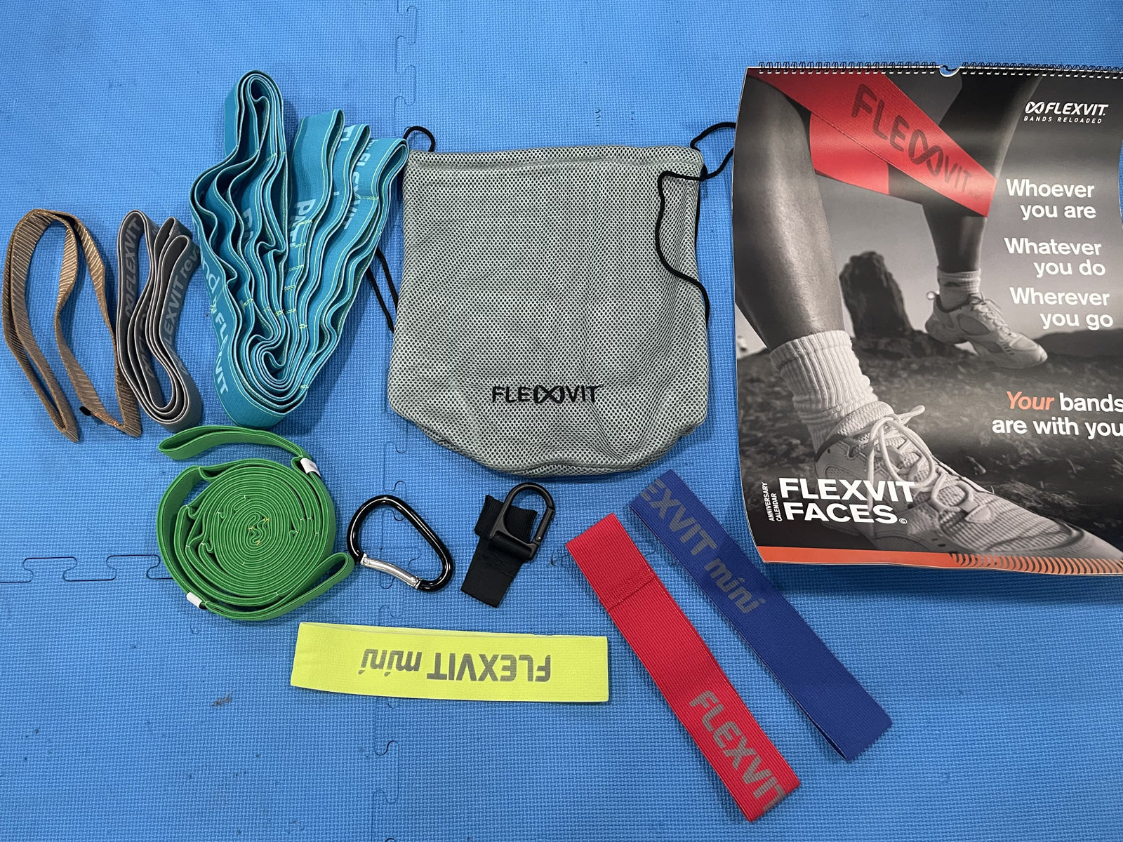
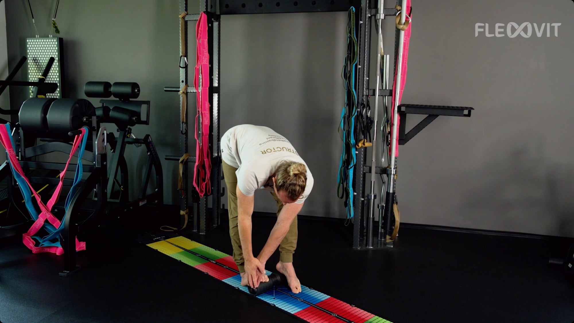
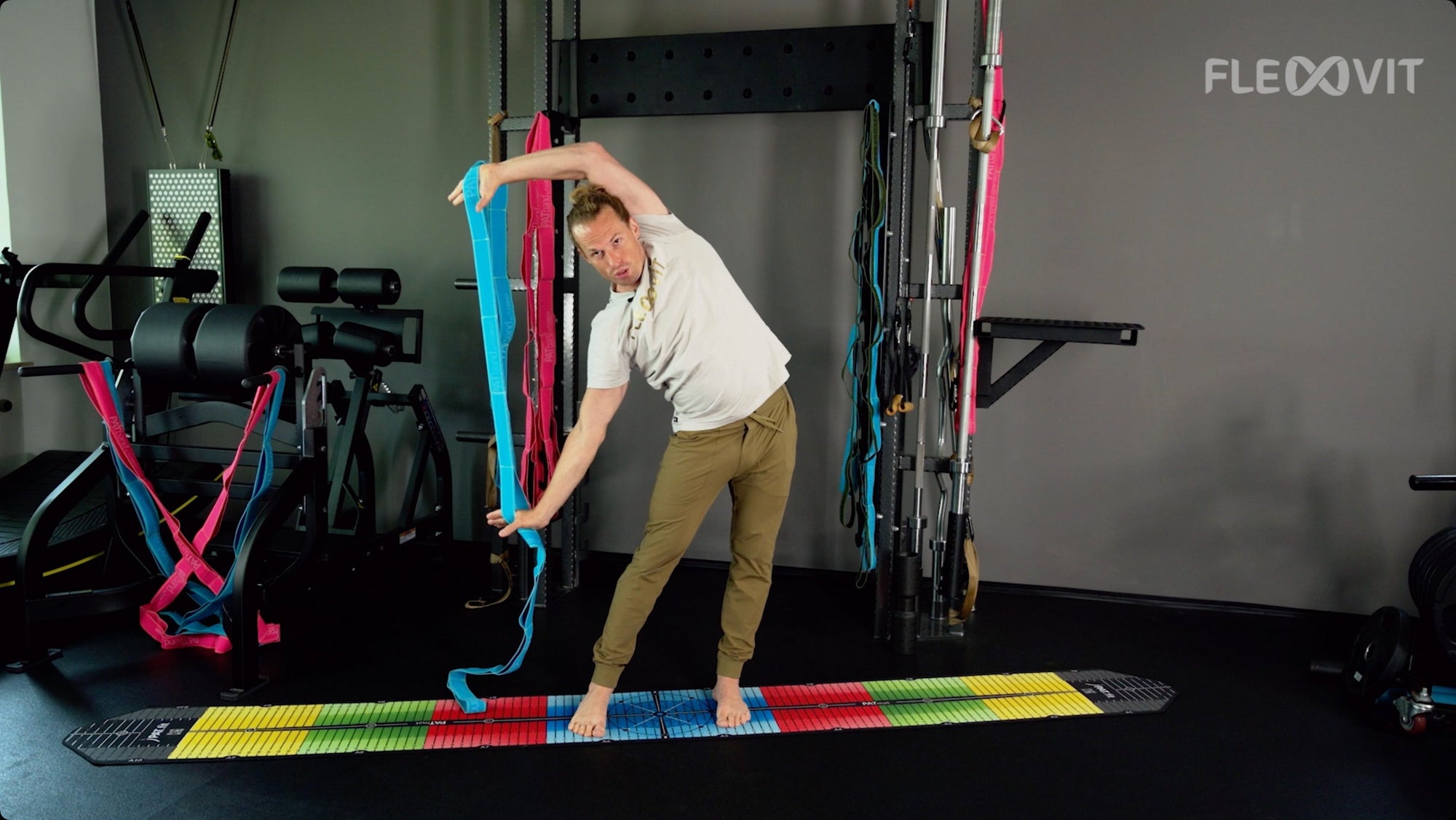
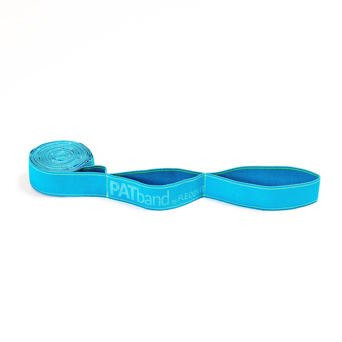
Let's look at some exercises that can be performed with elastic fitness bands (FLEXVIT ®). This can prevent ankle sprains and strengthen the muscle surrounding the ankle joint.
Fitness Band Exercises
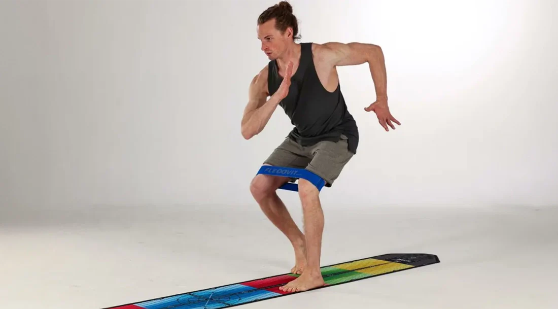
Exercise 1: Hip abduction and ankle eversion
Works the gluteus medius and peroneus muscle, which has been shown to be essential for rehabilitation after an ankle sprain or unstable ankle. It also prevents possible future ankle injuries.
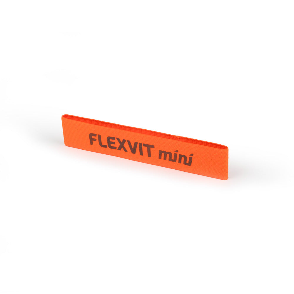
Order our FLEXVIT Mini now!
Exercise 2: Isolated ankle eversion
The Peroneus Muscles.
Exercise 3: Proprioception + Ankle Dorsiflexion Training
Sources:
Gibboney MD, Dreyer MA. Lateral ankle instability. [Updated 2020 Jul 6]. In: StatPearls [web]. Treasure Island (FL): StatPearls Publishing; 2020 Jan
Distributed under the terms of the Creative Commons Attribution 4.0 International License .
About the authors 
Antoine Fréchaud (left) and Nathan Touati (right) run NeuroXtrain, a website specializing in writing articles and various content on sports science, athlete rehabilitation, performance and new technologies for athlete health.
You can find more articles from NeuroXtrain on our blog or online.
Practical content such as videos on specific rehabilitation/prevention/strengthening exercises and much more:
Instagram | Facebook | LinkedIn

DISCLAIMER: This content (the description, images and videos) does not constitute medical advice or treatment plan and is intended for general educational and demonstration purposes only. This content should not be used to self-diagnose or self-treat any health, medical, or physical condition. This content cannot replace professional advice or a doctor's visit. Before exercises are used to treat specific complaints, a doctor or physiotherapist should always be consulted to be on the safe side.


